Advanced pre-operative and intra-operative image analysis with Artificial Intelligence
With market-leading capabilities in lung anatomical segmentation and advanced lung quantification, Thirona’s automatic analysis of chest CT and CBCT enable innovation along the complete clinical intervention pathway.
Target identification and treatment selection
Quantification of abnormalities for eligibility verification
Navigation planning
and decision-making
Precise visualization of diseases areas and target navigation assistance
Intervention
transbronchial and surgery
Intra-operative guidance
and decision-making support
Post-procedural
assessment
Post-treatment visualization of anatomy and disease quantification
High precision. Consistent performance.
Our modular AI platform LungQ™ contains robust high-performance algorithms, trained on a wide range of disease-specific datasets to deliver consistent and reproducible results.
The artificial intelligence-based quantitative analysis of Chest CT, include:
- Anatomical measures such as: volumes of each anatomical structure, parenchymal density evaluation, bronchial and vascular dimensions, bronchus-artery analysis (BA), fissure completeness, artery-vein phenotyping (AVX), etc.
- Disease-related severity and distribution assessment for: emphysema, air trapping, bronchial wall thickening, bronchial and vascular dropout, covid-19 infection, bronchiectasis, fibrosis, mucous impaction, atelectasis, CT-approximated perfusion (PXT) and ventilation defects, pulmonary vascular enlargement, etc.
Thirona’s advanced capabilities for anatomical segmentation of the lungs, lobes, pulmonary (sub)segments, interlobar fissures, airways and anatomical branches, pulmonary arteries and veins allow for precise exploration of local lung pathologies prior to intervention, during the procedure and post treatment.
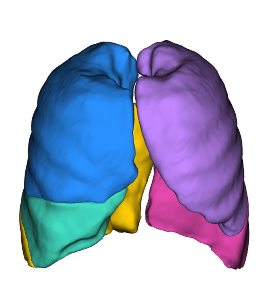
Anatomical segmentation
Lobar boundaries
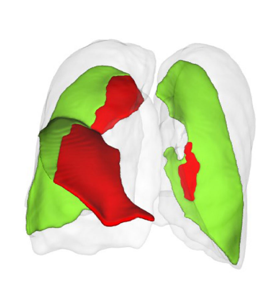
Anatomical quantification
Fissure completeness
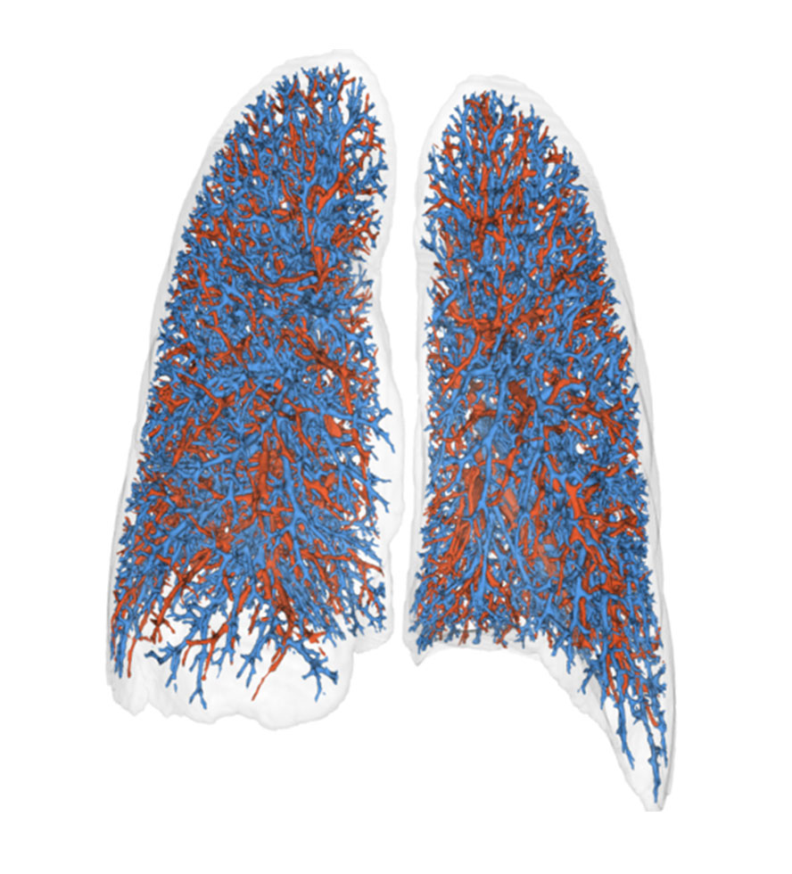
Anatomical segmentation
Artery-vein analysis
Disease severity assessment
Bronchiectasis
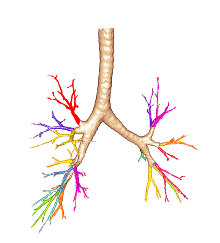
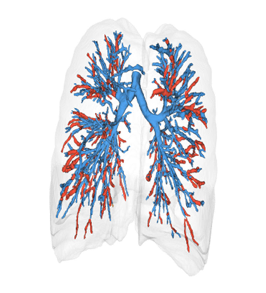
Anatomical segmentation
Anatomical airway generations
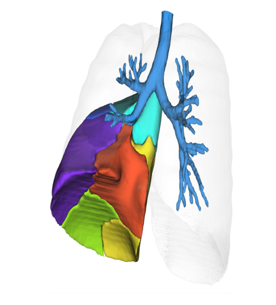
Anatomical segmentation
Segmental boundaries
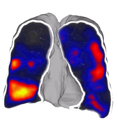
Anatomical quantification
Approximated perfusion (PXT)
Co-innovating for less invasive care, from concept exploration to clinical implementation
By partnering with leading technology players in the MedTech industry and clinical research institutions, we jointly translate the latest science and AI technology into certified clinical end-products that aid clinicians in advancing personalized treatment of lung patients.
The AI-based capabilities in pre-operative and intra-operative imaging have the potential to significantly enhance the precision, accuracy, and efficiency of (robotic) bronchoscopic and surgical lung interventions, enabling easier-to-perform and less invasive procedures.
With our extended domain expertise and proven track record in supporting medical innovations in interventional pulmonology, from proof-of-concept to clinical trials to clinical implementation, we can effectively tailor our services to specific project needs and challenges.
Why Thirona?
Market-proven co-innovation track record
State-of-the-art lung quantification AI platform
Unique domain expertise in lung diseases
Robust quality assurance methodology
LungQ 3.0.0 clinical software package
Approved for clinical use in Europe and USA
The Thirona’s LungQ v3.0.0 software produces CT values for pulmonary tissue, providing quantitative support for diagnosis and follow-up examinations. LungQ can be used to support the physician in the diagnosis, evaluation and documentation of pulmonary tissue images (e.g. abnormalities) from CT thoracic datasets. The analysis provided includes segmentation and isolation of sub-compartments, volumetric analysis, density evaluations, fissure evaluation and reporting tools.
LungQ v3.0.0 clinical software is FDA 510(k) cleared and a certified as a class IIb medical device CE marked (CE 0344) under the European Medical Devices Regulation (EU-MDR 217/745). The CE certified version provides additional analysis of tissue destruction and airway and vessels evaluation.
")