This blog provides a brief summary of a recent study “Children with severe asthma have substantial structural airway changes on computed tomography” by Wytse B. van den Bosch et al. (ERS Journal, Jan’2024), highlighting key insights and potential impact on treatment of the disease.
Around 10% of children with asthma are classified as severe. This means they continue to suffer from severe asthma symptoms despite adequate anti-asthma therapy. The reason why is mostly not clear. In many of these cases a lung CT is made to evaluate whether there is damage to the lung structure and to exclude alternative diagnosis. The evaluation performed by expert radiologists and pediatric pulmonologists is traditionally done by eyeballing. Unfortunately, this visual interpretation of the images is insensitive to thoroughly assess the condition of the hundreds of bronchi that are captured on a CT scan. Typically, this evaluation is based on around 20-30 mostly larger bronchi which can be assessed visually. The more experienced the specialist, the more accurate the assessment.
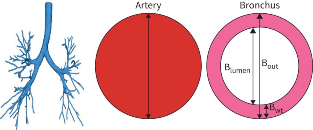
Thanks to the development of fully automatic image analysis systems enabled by artificial intelligence, it has become now possible to measure the dimensions of almost all airways and blood vessels on a CT scan, with great precision and accuracy.
Seeing the ‘unseen’, measuring the ‘unmeasurable’
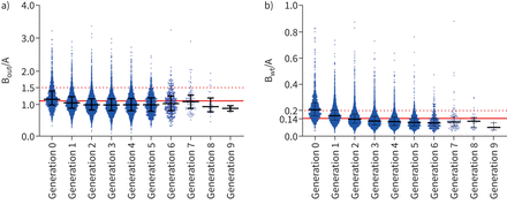
Unexpectedly, it was shown that almost all these children have substantial abnormalities of airways, with bronchial dilatation (defined as bronchiectasis, currently thought to be irreversible) detected in one third of the patients. These findings are of great value for enhancing our understanding of severe asthma and for learning more on the development of bronchiectasis disease as diagnosed in adults. The ability of artificial intelligence to assess changes in bronchial dilatation and bronchial wall thickening with high sensitivity, can potentially help uncover new insights on the development of permanent airway damage.
Furthermore, it opens the possibility to use lung CT images to evaluate the effectiveness of novel therapies targeted at improving the condition of the airways and even potentially preventing the development of irreversible impairment.
Finally, the study underlines the importance of routinely including a lung CT with automatic analysis of the images to ensure objective and sensitive assessment of airway damage in patients with severe asthma and to personalize treatment and monitoring.
The ability of artificial intelligence to assess changes in bronchial dilatation and bronchial wall thickening with high sensitivity, can potentially help uncover new insights on the development of permanent airway damage