MedicalVR has integrated Thirona’s chest CT analysis software in their virtual reality application PulmoVR. It provides surgeons with an interactive 3D visualization of the lungs for pre-operative planning. Patient-specific anatomical labels and segmentation are added by our CT analysis software, allowing for more targeted surgery and sparing healthy lung tissue.
Surgeons navigating patients’ lungs in virtual reality: it may seem futuristic, but is reality in Erasmus Medical Center in Rotterdam. Together with our partner MedicalVR, the cardio-thoracic surgery department of the Erasmus MC has started a pilot with 20 patients, using virtual reality in preparation for complex lung surgery. Thirona’s chest CT analysis is at the core of the application, using artificial intelligence to distinguish anatomical features and providing detailed segmentation of the lungs.
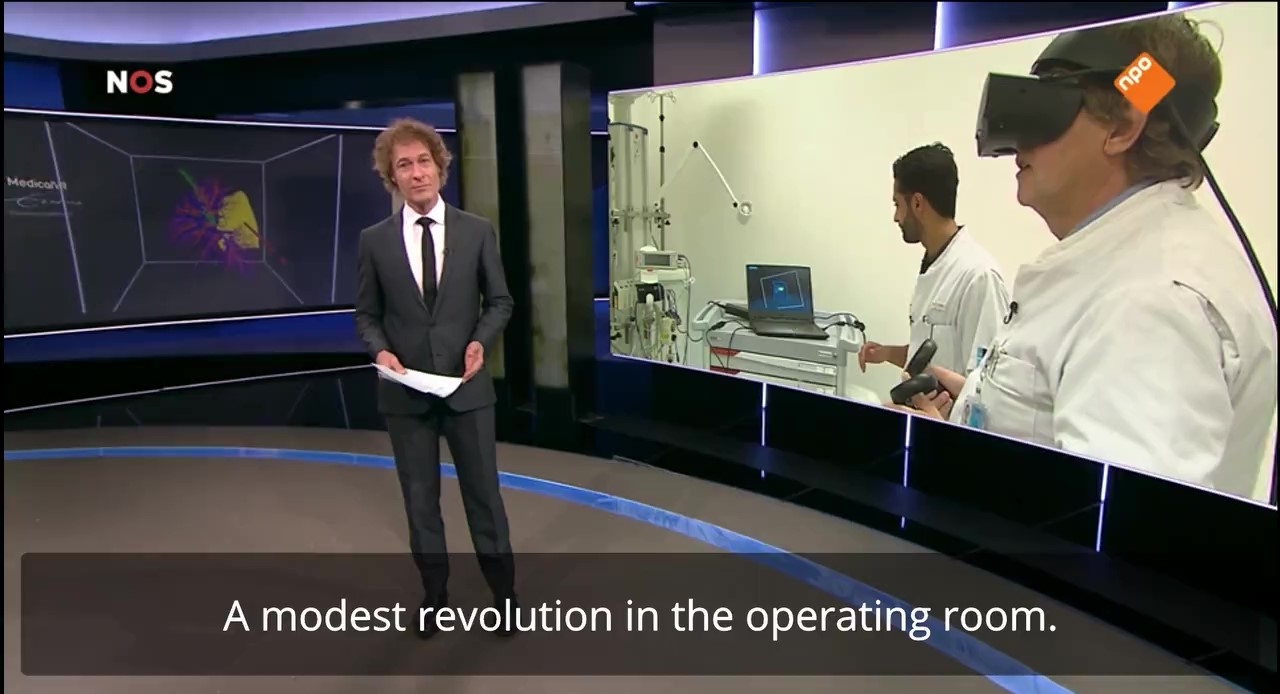
The project was featured on the Dutch national news channel NOS last weekend, where the VR application was mentioned as “a modest revolution in the operating room”. What makes the 3D virtual reality model so special, is that it lets surgeons see the location of tumors and other abnormalities in the lungs with much greater precision than is possible on regular CT images. This allows for more targeted surgery to accurately remove a tumor from a certain part of the lung. The result: more healthy lung tissue and breathing capacity is spared.
Thirona’s analysis of the thoracic CT scans not only labels the major anatomical structures of the lungs, such as lobes, airways and blood vessels. It also provides automated segmentation of the lungs into sub-lobar segments, which is especially valuable for the precise localization of lung abnormalities The analysis is based on state-of-the-art deep learning technology, like all Thirona’s software solutions.
Watch the full news item here: